

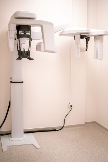
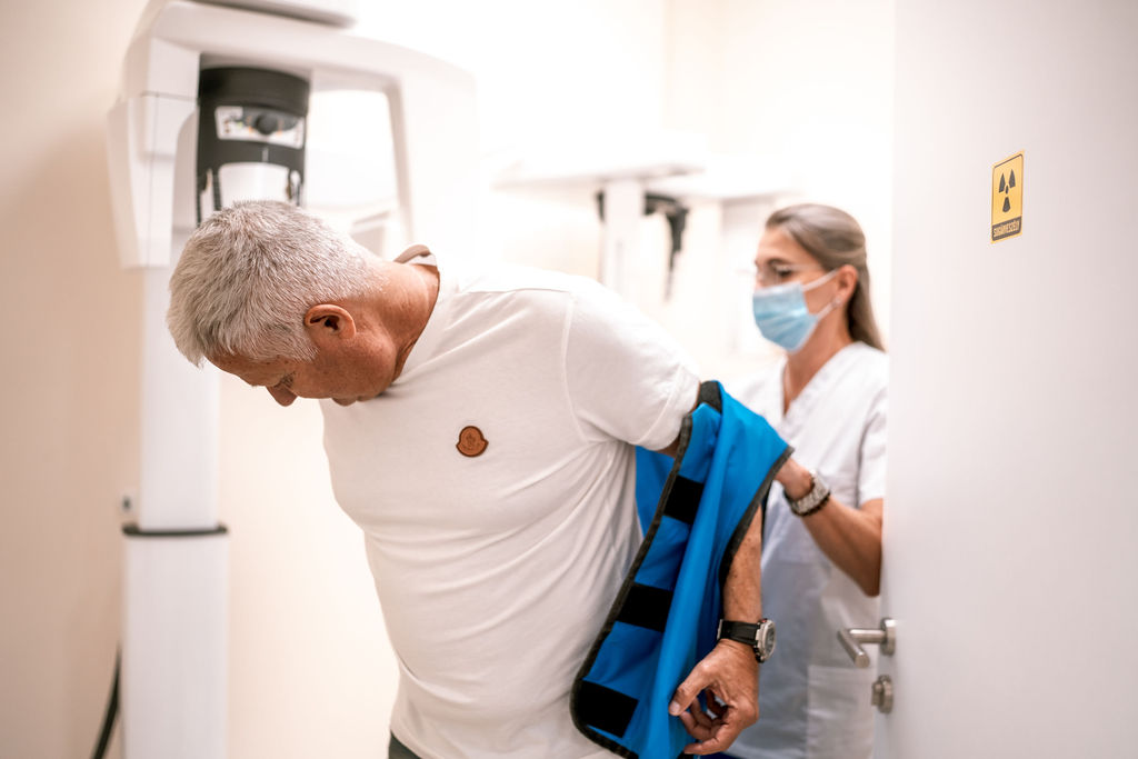
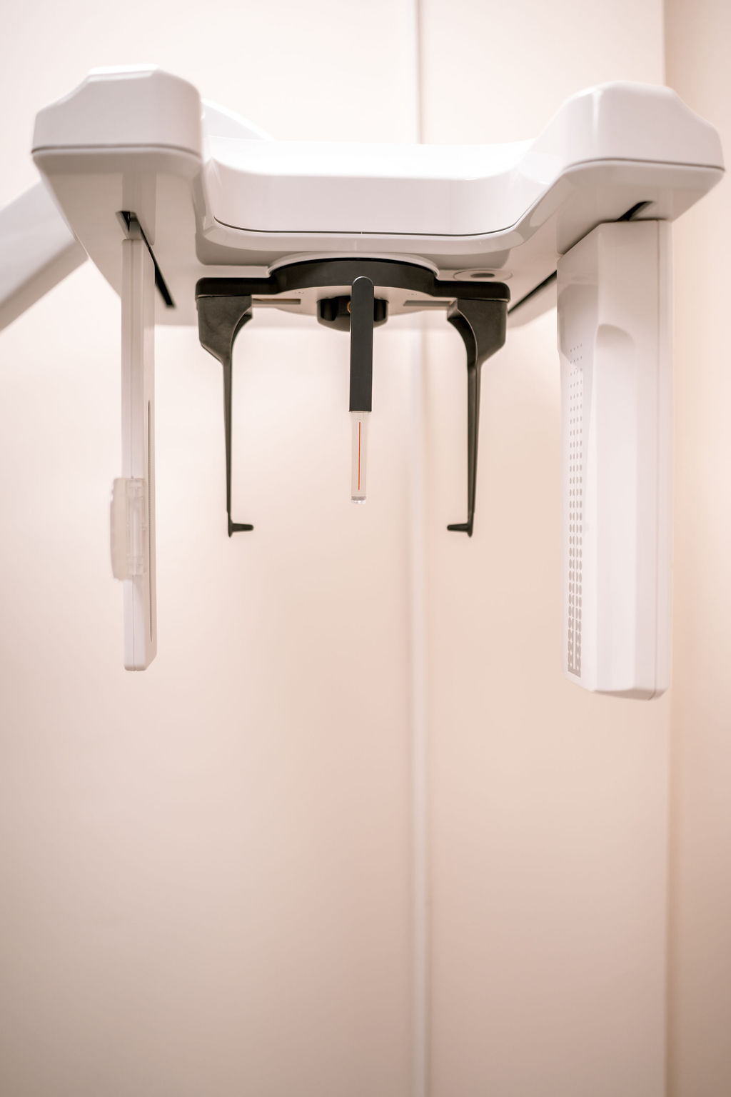
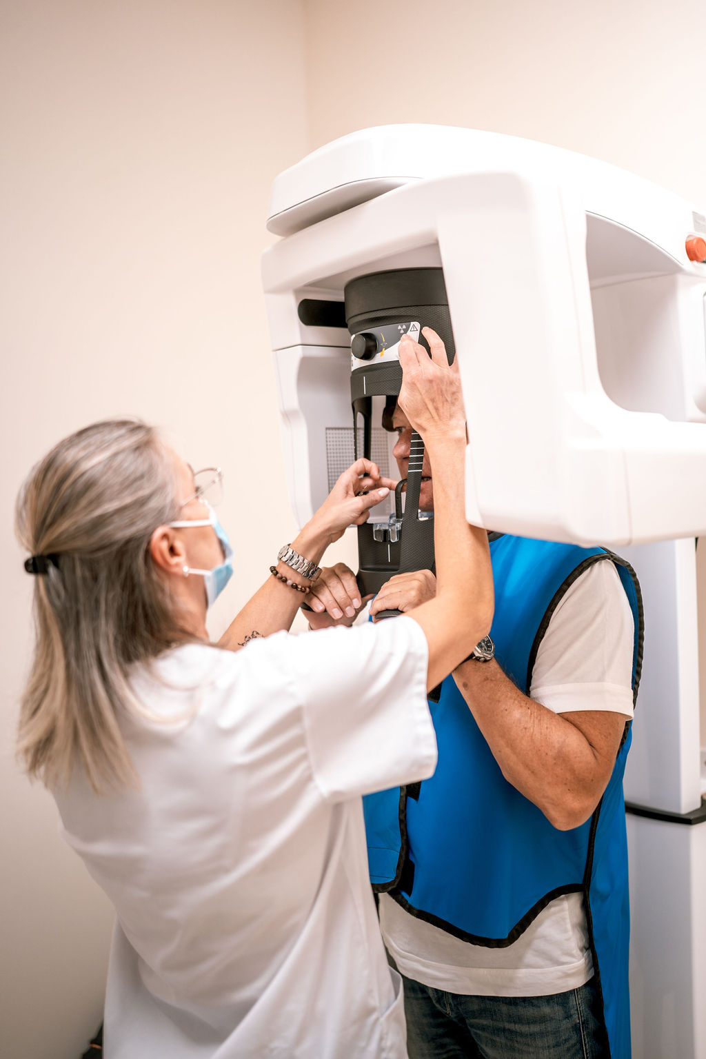
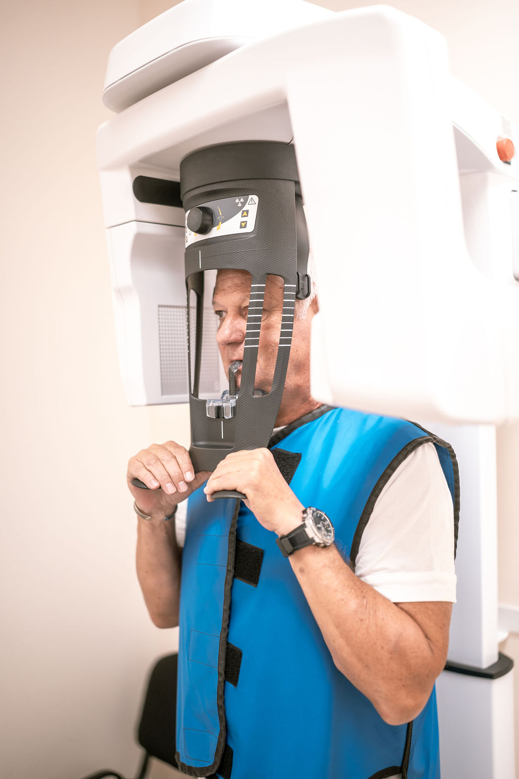
The foundation of modern dental diagnostics is accurate, detailed imaging, which enables the early detection of hidden abnormalities and precise treatment planning. At our practice, we have state-of-the-art CBCT (3D dental CT) equipment, providing high-resolution, three-dimensional images of the structures of the oral cavity and jawbones.
In addition to CBCT, we can also take targeted images covering smaller areas, allowing us to perform examinations with the lowest radiation dose necessary. Digital X-ray is a fast, accurate, and low-dose procedure that forms the basis of everyday dental diagnostics.
Detailed imaging is particularly important when planning implant treatments, root canal treatments, and surgical procedures, as it enables maximum safety and predictability throughout the treatment process.
For examining the teeth and the surrounding bone structure, we use a computer-connected dental X-ray system, in which conventional film is replaced by a special sensor connected to a computer.
The resulting image is displayed immediately on a monitor positioned in front of the patient, allowing the dentist to show any pathological changes on an enlarged image.
Another major advantage is that the radiation dose is only a fraction of that used by conventional X-ray equipment.
The X-ray images, together with the dental chart and intraoral camera images, are stored on the computer and can be retrieved at any time in the future, allowing the original condition to be compared with the condition after treatment and healing.


4.9/5
Google Rating
20+
Years of experience
10.000+
Happy patients